
Plaque psoriasis
Recognise a sharply demarcated erythematous plaque with adherent silvery scale on an extensor surface.
Updated: 2026-09-13
Written for clinicians and medical students
This section is a teaching reference for doctors, residents and medical students. It is not patient guidance and not a substitute for clinical examination, and it must not be used for self treatment.
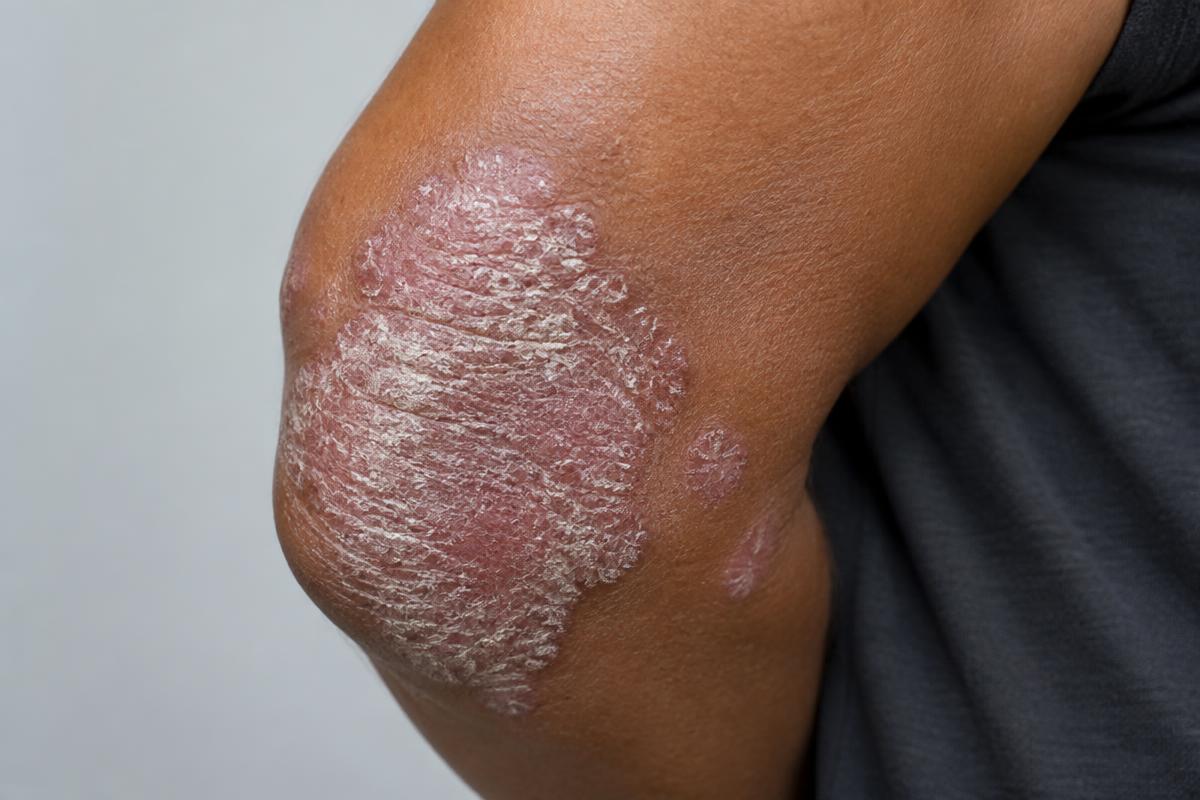
AI-generated image for education only. It is not a real patient photograph or pathology specimen.
Typical teaching pattern: a well-demarcated plaque with thick silvery scale on the elbow.
Clinical clues
- Look for symmetry and involvement of the scalp, umbilicus, natal cleft, nails and other extensor surfaces.[1]
- Scale may be less obvious on flexural sites. Erythema can appear violaceous or dark brown in richly pigmented skin.[1]
- Important mimics include tinea corporis, nummular dermatitis, lichen simplex and cutaneous T-cell lymphoma.
Confirm and assess
- Diagnosis is usually clinical. Perform KOH microscopy when an annular lesion or asymmetric scale raises concern for dermatophyte infection.
- Assess body surface area, difficult sites, symptoms, quality of life, joint symptoms and cardiometabolic risk before choosing treatment.[2]