
Tinea corporis
Identify an annular plaque with peripheral scale, an advancing border and relative central clearing.
Updated: 2026-09-13
Written for clinicians and medical students
This section is a teaching reference for doctors, residents and medical students. It is not patient guidance and not a substitute for clinical examination, and it must not be used for self treatment.
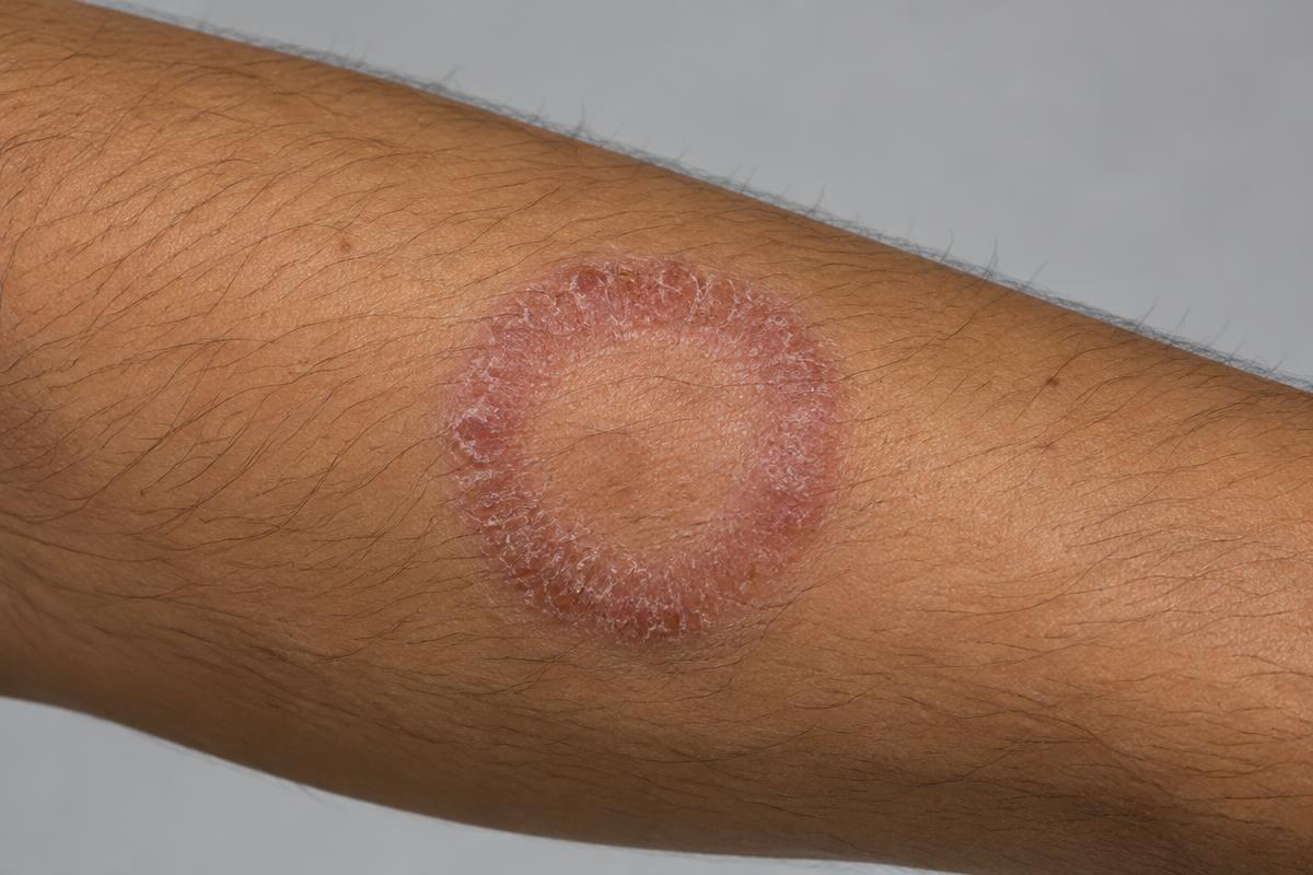
AI-generated image for education only. It is not a real patient photograph or pathology specimen.
Teaching pattern: annular erythema with a raised scaly active edge and central clearing.
Clinical clues
- The active edge is usually more erythematous and scaly than the centre. Lesions enlarge centrifugally and are often itchy.[1]
- Ask about infected household contacts, pets, contact sport, occlusive clothing and topical corticosteroid use.
- Topical corticosteroids can suppress scale and distort the border, producing tinea incognito.[1]
Confirm and differentiate
- Scrape the active border for KOH microscopy before treatment when the diagnosis is uncertain.[2]
- Consider nummular dermatitis, pityriasis rosea, granuloma annulare, psoriasis and erythema migrans according to morphology and history.