
Vitiligo
Recognise acquired, sharply demarcated depigmented macules and patches without surface change.
Updated: 2026-09-13
Written for clinicians and medical students
This section is a teaching reference for doctors, residents and medical students. It is not patient guidance and not a substitute for clinical examination, and it must not be used for self treatment.
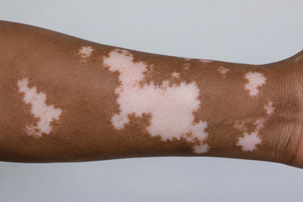
AI-generated image for education only. It is not a real patient photograph or pathology specimen.
Teaching pattern: sharply demarcated depigmented patches without scale or textural change.
Clinical clues
- Common sites include the face, hands, body folds, genital skin and areas of friction. Leukotrichia can occur.[1]
- A Wood lamp accentuates depigmentation and helps define extent, especially in lighter skin.[1]
- Distinguish complete depigmentation from post-inflammatory hypopigmentation, pityriasis alba, tinea versicolor and chemical leukoderma.
Assessment
- Document distribution, body surface area, activity, new lesions and impact on quality of life.
- Review symptoms and history for associated autoimmune disease. Testing should be guided by clinical context rather than performed indiscriminately.[2]