Basal cell carcinoma histopathology
Identify basaloid tumour islands with peripheral palisading and stromal retraction.
Updated: 2026-09-13
Written for clinicians and medical students
This section is a teaching reference for doctors, residents and medical students. It is not patient guidance and not a substitute for clinical examination, and it must not be used for self treatment.
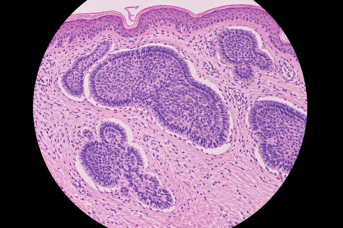
AI-generated image for education only. It is not a real patient photograph or pathology specimen.
Synthetic H&E teaching image. Review basaloid nests, peripheral palisading and retraction clefts.
Systematic description
- Nests and cords of basaloid cells arise from the epidermis or follicular epithelium and extend into the dermis.[1]
- Peripheral nuclear palisading and cleft-like retraction between tumour and mucinous stroma are classic clues.[1]
- Report the histological subtype, depth, perineural or lymphovascular invasion, and margin status when applicable.[2]
Differential and caveat
- Trichoepithelioma, trichoblastoma, basaloid squamous carcinoma and microcystic adnexal carcinoma may enter the differential.
- Infiltrative, micronodular, morpheaform and basosquamous patterns carry greater recurrence risk than superficial or nodular patterns.[2]