
Lichen planus histopathology
Recognise a lichenoid interface dermatitis with a dense band of lymphocytes at the dermoepidermal junction.
Updated: 2026-09-13
Written for clinicians and medical students
This section is a teaching reference for doctors, residents and medical students. It is not patient guidance and not a substitute for clinical examination, and it must not be used for self treatment.
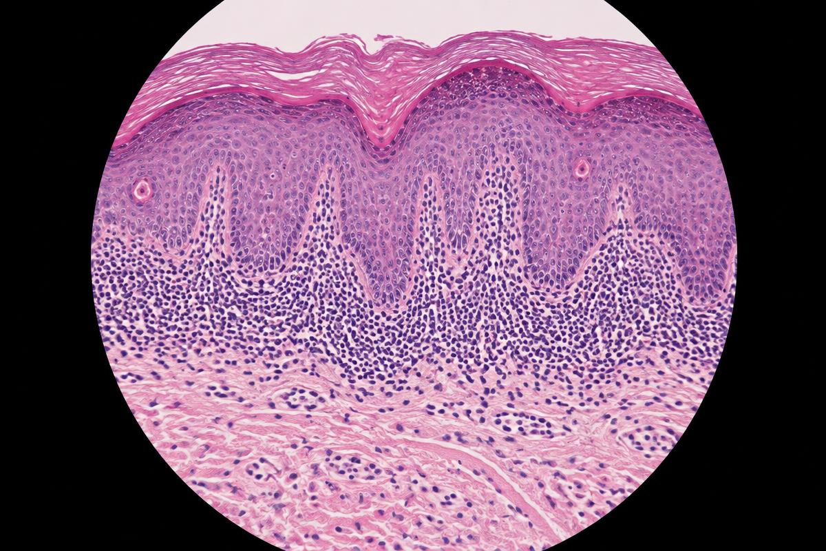
AI-generated image for education only. It is not a real patient photograph or pathology specimen.
Synthetic H&E teaching image. Note the band-like interface infiltrate and irregular epidermal contour.
Systematic description
- Compact orthokeratosis, wedge-shaped hypergranulosis and irregular acanthosis with pointed or sawtooth rete ridges.[1]
- A dense band-like lymphocytic infiltrate obscures the dermoepidermal junction, with basal vacuolar change and apoptotic keratinocytes.[1]
- Pigment incontinence and melanophages are common, especially in older lesions and richly pigmented skin.
Differential and caveat
- Consider lichenoid drug eruption, lupus erythematosus, graft-versus-host disease and lichenoid keratosis.
- Eosinophils, parakeratosis and a deeper perivascular infiltrate can favour a lichenoid drug reaction, but medication history remains essential.[2]