
Psoriasis histopathology
Read the psoriasiform reaction pattern through regular acanthosis, parakeratosis and neutrophils.
Updated: 2026-09-13
Written for clinicians and medical students
This section is a teaching reference for doctors, residents and medical students. It is not patient guidance and not a substitute for clinical examination, and it must not be used for self treatment.
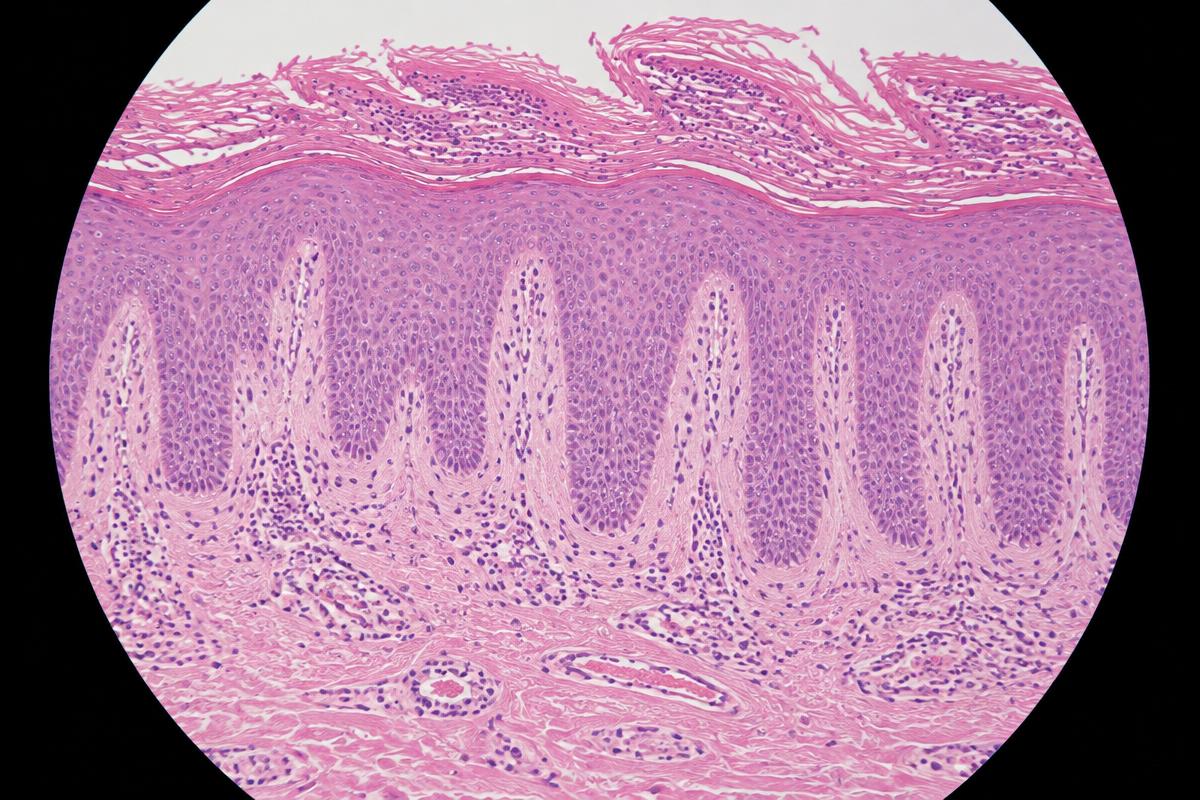
AI-generated image for education only. It is not a real patient photograph or pathology specimen.
Synthetic H&E teaching image. Review the regular rete ridge elongation, parakeratosis and superficial inflammation.
Systematic description
- Regular psoriasiform epidermal hyperplasia with elongated rete ridges and thinning over dermal papillae.[1]
- Confluent parakeratosis, reduced or absent granular layer, and neutrophils in the stratum corneum or epidermis.[1]
- Dilated papillary dermal capillaries and a superficial perivascular lymphocytic infiltrate support the pattern.
Differential and caveat
- Seborrhoeic dermatitis, chronic spongiotic dermatitis, pityriasis rubra pilaris and dermatophyte infection can be psoriasiform.
- Early, treated or atypical lesions may lack the full pattern. Clinicopathological correlation and fungal stains can be decisive.[2]