Surgical excision of skin lesions: margins, closure and scars
Excision removes the lesion and provides a specimen for the laboratory. That second point is what makes it the definitive procedure for anything suspicious.
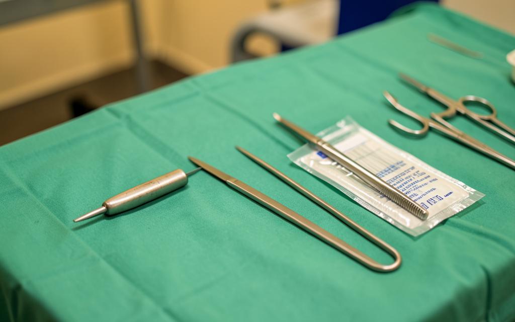
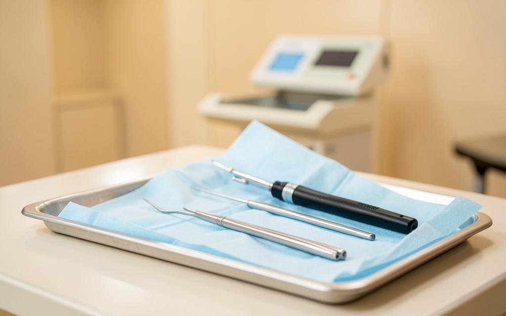
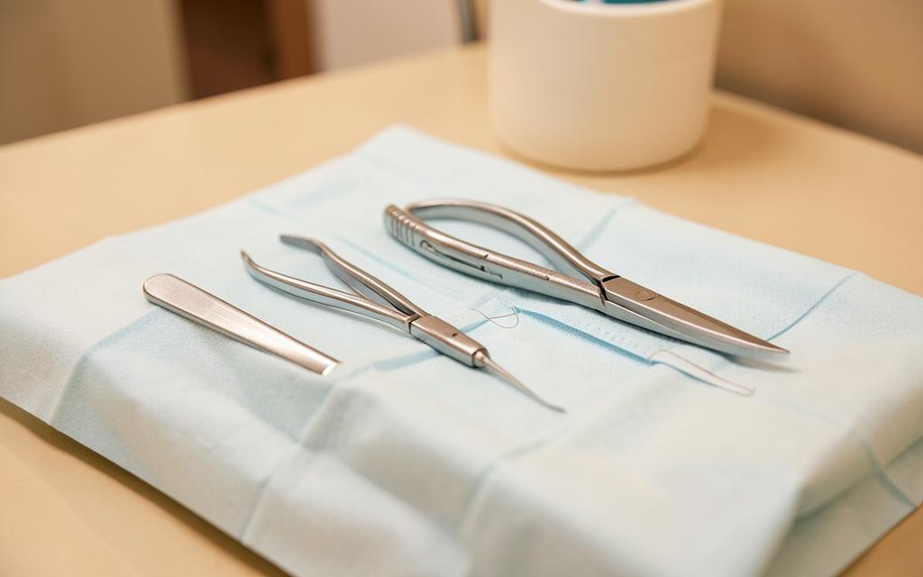
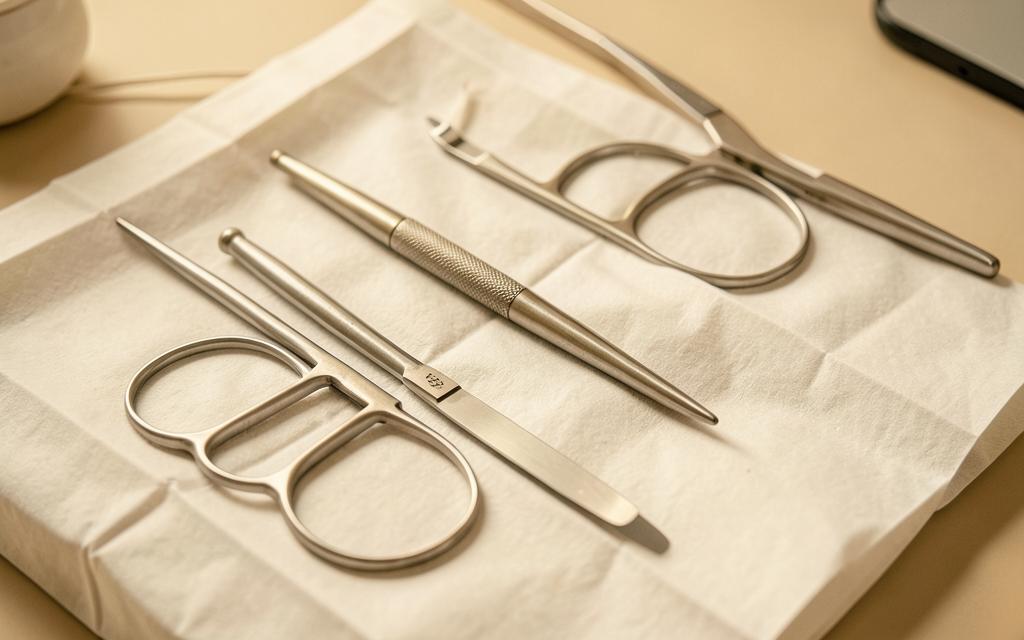
Excision under local anaesthesia is the workhorse of dermatologic surgery. An ellipse of skin containing the lesion and a planned margin is removed, the defect is closed in layers, and the specimen is sent for histopathology. Unlike destructive methods, excision produces a diagnosis as well as a treatment [1].
Key points
- Any lesion suspicious for skin cancer should be excised or biopsied, never destroyed blindly
- Margins depend on diagnosis, from a narrow margin for a benign mole to defined margins for melanoma
- The ellipse is oriented along relaxed skin tension lines to minimise the scar
- Histopathology confirms the diagnosis and whether the excision was complete
- Every excision leaves a scar, and scar quality depends on site, tension and aftercare
Planning the excision
The lesion is marked with the naked eye and with dermoscopy, and the margin is decided from the working diagnosis. A benign mole removed for cosmetic reasons needs only the lesion itself, while a basal cell carcinoma typically needs a several millimetre margin and a melanoma has margins defined by tumour thickness in national guidance [2].
The long axis is placed along relaxed skin tension lines so that the scar sits within a natural crease and closure is under minimal tension. Length is usually about three times the width so the wound closes flat without standing cones at the ends.
The procedure
The skin is cleaned and local anaesthetic, usually lidocaine with adrenaline, is injected. Adrenaline reduces bleeding and prolongs anaesthesia. After a few minutes the area is completely numb, and the patient feels pressure and movement but not pain.
The ellipse is incised to the required depth, the specimen is lifted with a skin hook to avoid crush artefact, haemostasis is achieved, and the wound is closed with deep absorbable sutures followed by fine surface sutures. Most excisions take twenty to forty minutes.
- Face: fine sutures removed at five to seven days
- Trunk and limbs: sutures removed at ten to fourteen days
- Back and shoulders: highest tension, widest scars, longest support with tape
- Lower leg: slowest healing, particularly in diabetes or venous disease
Histopathology and what the report means
The specimen is fixed, processed and examined by a pathologist. The report confirms the diagnosis and states whether the lesion was completely excised and how close it came to the margins. If a skin cancer is incompletely excised, further surgery is usually recommended.
This is precisely why a changing mole should not be treated with laser or cryotherapy. Destroying tissue eliminates the chance to examine it, and a melanoma treated that way can recur deeper with a delayed diagnosis [2].
Aftercare and scars
Keep the wound dry for forty eight hours, then follow the dressing plan given. Avoid stretching the area and heavy lifting for two weeks, longer for the back. Bleeding, spreading redness, increasing pain or discharge should be reviewed promptly.
A scar remains red and firm for two to three months and softens over a year. Silicone gel or sheeting, taping and sun protection improve the final appearance. Patients with a keloid tendency, particularly on the chest, shoulders and earlobes, are counselled before surgery and may need preventive intralesional steroid.
Frequently asked questions
Will the mole grow back?
A completely excised mole does not return. Partial removal, for example by shave, can leave pigment that regrows, which is why complete excision is preferred for anything atypical.
Does removing a mole make cancer spread?
No. This is a persistent myth. Proper excision of a melanoma is the treatment, and delay is what worsens outcomes.
How big will the scar be?
Longer than the lesion, because the ellipse must be several times longer than it is wide to close flat. A five millimetre lesion typically leaves a scar of around two centimetres.
References
- [1] Kimyai Asadi A, Goldberg LH. Basics of dermatologic surgery: excisional technique and closure. Source
- [2] Swetter SM, Tsao H, et al. Guidelines of care for the management of primary cutaneous melanoma. Journal of the American Academy of Dermatology. Source
- [3] National Institute for Health and Care Excellence guidance. Source
This content is general medical education and does not replace a personal consultation. Any prescription must be coordinated with your local doctor.
Is this procedure right for you?
Book a video consultation to discuss your case, or a clinic visit in Baghdad or Karbala.