Dermoscopy and mole mapping: catching skin cancer earlier
A dermatoscope reveals structures invisible to the eye. In trained hands it raises melanoma detection accuracy and reduces unnecessary surgery.
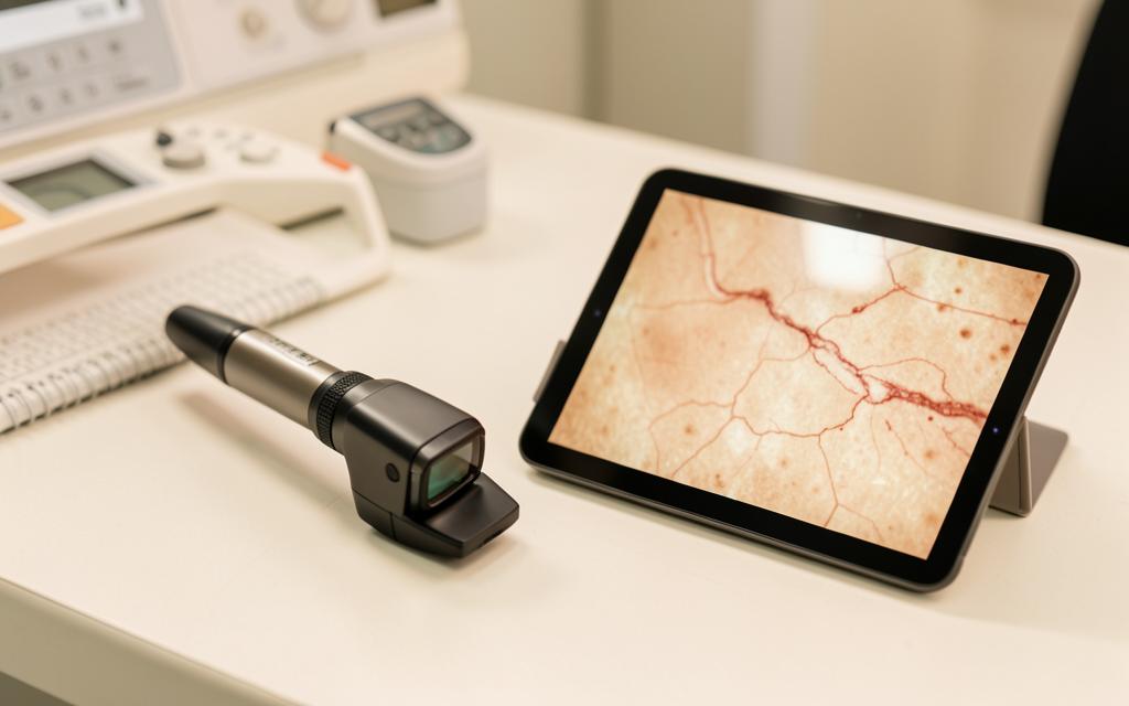
Dermoscopy uses magnification with polarised or immersion light to see below the stratum corneum and visualise pigment networks, vascular patterns and structures that distinguish benign from malignant lesions. Meta analyses show it improves diagnostic accuracy for melanoma compared with naked eye examination when performed by trained clinicians [1].
Key points
- Improves melanoma detection accuracy and reduces unnecessary excisions
- Also used for hair, nails, scabies, warts and inflammatory conditions
- Digital monitoring detects change over time in patients with many moles
- Full body photography suits high risk patients with atypical mole syndrome
- A changing lesion is excised regardless of a reassuring dermoscopic appearance
What dermoscopy shows
Beneath the surface, benign naevi show organised, symmetrical patterns: a regular pigment network, uniform globules or a homogeneous blue grey structure. Melanoma tends to show asymmetry of structure and colour, an atypical network, irregular streaks, blue white veil and atypical vessels.
Basal cell carcinoma has its own signature of arborising vessels, leaf like areas and blue grey ovoid nests, which allows confident diagnosis in many cases before biopsy.
- Pigmented lesions: distinguishing naevus, seborrhoeic keratosis, lentigo and melanoma
- Non pigmented tumours: vascular patterns of basal and squamous cell carcinoma
- Trichoscopy: alopecia areata, androgenetic alopecia and scarring alopecias
- Onychoscopy: nail pigmentation and splinter haemorrhages
- Entodermoscopy: scabies mites, lice and molluscum
Mole mapping and digital monitoring
Sequential digital dermoscopy photographs individual lesions and compares them at intervals, usually three and then twelve months. Change over time is one of the most reliable signals of early melanoma, particularly for lesions that look mildly atypical but not clearly malignant.
Total body photography records the whole skin surface so that new lesions can be identified at follow up. Together these techniques allow high risk patients to be monitored closely while avoiding excision of every atypical looking mole [2].
Who should be monitored
Monitoring is aimed at people at elevated risk: a personal history of melanoma, a strong family history, more than fifty naevi or multiple atypical naevi, a history of significant sunburn or tanning bed use, immunosuppression, and fair skin that burns easily.
In Iraq, where most patients have Fitzpatrick types three to five, melanoma is less common but acral lentiginous melanoma on the soles, palms and nails is relatively more frequent and is often diagnosed late. Any changing lesion on the sole or a new pigmented band on a nail deserves examination.
Limits
Dermoscopy is operator dependent. In untrained hands accuracy is no better, and sometimes worse, than the naked eye, so training matters more than the device.
Featureless or amelanotic melanoma can appear bland. The rule remains that a lesion which is changing, growing, bleeding or simply different from the patient other moles is investigated regardless of a reassuring dermoscopic image.
Frequently asked questions
Is a mole check painful or invasive?
Not at all. The dermatoscope touches the skin or hovers above it. A full skin examination takes fifteen to twenty minutes.
How often should I have a check?
For most people, only when a lesion changes. High risk patients are usually seen every six to twelve months.
Can a photograph sent online replace an examination?
A good quality photograph can triage urgency and identify obviously benign or clearly suspicious lesions, but full assessment of a pigmented lesion needs dermoscopy in person.
References
- [1] Vestergaard ME, Macaskill P, Holt PE, Menzies SW. Dermoscopy compared with naked eye examination for the diagnosis of primary melanoma: a meta analysis. British Journal of Dermatology. Source
- [2] Salerni G, Carrera C, et al. Benefits of total body photography and digital dermatoscopy in the early diagnosis of melanoma. Journal of the American Academy of Dermatology. Source
This content is general medical education and does not replace a personal consultation. Any prescription must be coordinated with your local doctor.
Is this procedure right for you?
Book a video consultation to discuss your case, or a clinic visit in Baghdad or Karbala.