Skin biopsy: how a diagnosis is actually made
When the eye and the dermatoscope are not enough, a few millimetres of skin under a microscope settle the question.
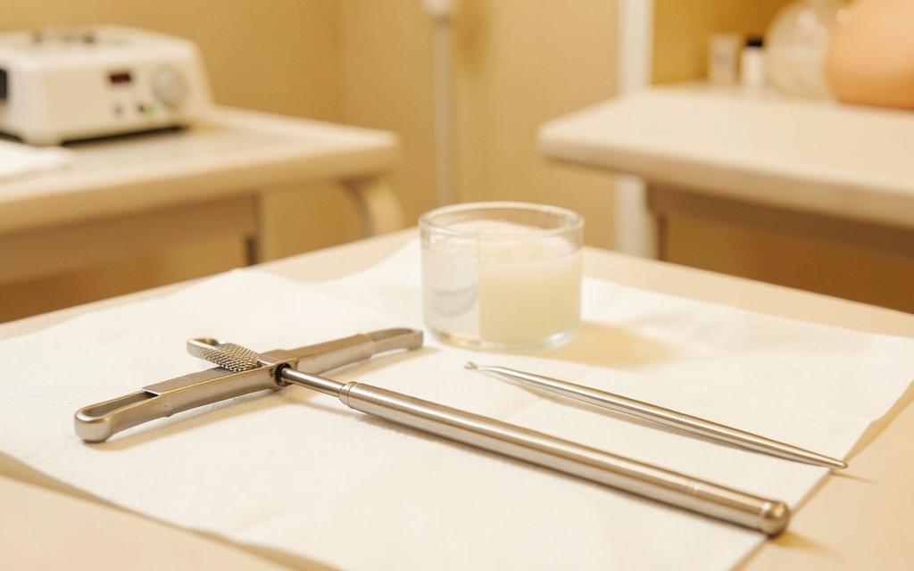
A skin biopsy takes a small sample for microscopic examination. It is the reference standard for diagnosing inflammatory skin disease, confirming skin cancer, investigating hair loss and identifying blistering disorders. Choosing the right biopsy type and site is as important as the procedure itself, because a poorly chosen sample can be uninformative [1].
Key points
- Punch biopsy gives a full thickness core and is the general purpose choice
- Shave biopsy suits raised lesions but should not be used where depth matters
- Excisional biopsy removes the whole lesion and is preferred for suspected melanoma
- Immunofluorescence for blistering disease needs a second sample in special transport medium
- Results typically take one to two weeks
Types of biopsy and when each is used
A punch biopsy uses a circular blade, usually three to five millimetres, to take a full thickness cylinder including epidermis, dermis and some subcutis. It is used for rashes, suspected lymphoma, granulomatous disease and most inflammatory conditions.
A shave biopsy removes a raised lesion at or just below the surface with a blade. It suits seborrhoeic keratoses, basal cell carcinomas planned for further treatment and benign raised lesions, but is avoided where measuring the depth of a tumour matters, as in melanoma, because a shave that transects the lesion base can make accurate staging impossible [2].
- Rash or inflammatory disease: punch from a fully developed, untreated lesion
- Suspected melanoma: complete excisional biopsy with a narrow margin
- Suspected basal cell carcinoma: punch or shave for diagnosis before definitive treatment
- Hair loss: two four millimetre punches from the active area, one for horizontal sections
- Blistering disease: lesional biopsy for histology plus perilesional biopsy for immunofluorescence
Choosing the site
The most informative site is a fresh, fully developed and untreated lesion. Biopsying a scratched, secondarily infected, or steroid treated lesion often produces a non specific report. For vasculitis, an early purpuric lesion less than forty eight hours old gives the best yield.
Cosmetic and functional considerations also matter: avoiding the lower leg where healing is slow, avoiding the central face where scars are conspicuous, and avoiding areas over tendons or joints.
The procedure
The site is cleaned and local anaesthetic is injected, which stings briefly. The punch is rotated through the skin, the core is lifted with forceps and cut free at the base, and the defect is closed with one or two sutures or allowed to heal on its own for small punches.
The whole process takes ten to fifteen minutes. The specimen goes into formalin, except immunofluorescence samples which require Michel medium or saline transport, so the clinical question must be stated on the request form before the biopsy is taken.
Understanding the report
A pathology report describes the architecture and cell types seen and gives a diagnosis, a differential, or sometimes a descriptive pattern such as spongiotic dermatitis or interface dermatitis, which must be correlated with the clinical picture.
A non diagnostic result does not mean the biopsy was wasted. It can exclude serious diagnoses and guide the next step, which may be a repeat biopsy from a different or more active site.
Frequently asked questions
Does a biopsy hurt?
The anaesthetic injection stings for a few seconds. After that you feel pressure only. There is mild soreness for a day or two.
Will it leave a scar?
Yes, usually a small round or linear mark that fades over months. Site selection takes visibility into account.
How long do results take?
Usually one to two weeks, longer if special stains or a second opinion are required.
References
- [1] Nischal U, Nischal KC, Khopkar U. Techniques of skin biopsy and practical considerations. Journal of Cutaneous and Aesthetic Surgery. Source
- [2] Swetter SM, Tsao H, et al. Guidelines of care for the management of primary cutaneous melanoma. Journal of the American Academy of Dermatology. Source
- [3] American Academy of Dermatology clinical guidelines. Source
This content is general medical education and does not replace a personal consultation. Any prescription must be coordinated with your local doctor.
Is this procedure right for you?
Book a video consultation to discuss your case, or a clinic visit in Baghdad or Karbala.