Wood's lamp examination: what ultraviolet light reveals about your skin
A simple handheld ultraviolet lamp in a darkened room can separate vitiligo from other pale patches, confirm certain fungal and bacterial infections, and show whether pigment is superficial or deep.
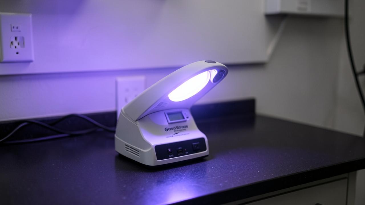
A Wood's lamp emits long wave ultraviolet light, around 365 nm, filtered through nickel oxide glass. Different skin pigments, fungi and bacteria absorb or fluoresce under that wavelength in characteristic ways [1]. The test takes under a minute, costs almost nothing, causes no discomfort, and frequently changes the diagnosis in the room. It is one of the few investigations in dermatology where the answer arrives before the patient leaves the chair.
Key points
- Painless, takes seconds, and needs no preparation beyond a darkened room
- Vitiligo glows bright chalky white with sharp borders; most other pale patches do not [1]
- Coral red fluorescence in a body fold points to erythrasma, a bacterial not fungal infection [2]
- It shows whether melasma pigment is mainly epidermal or dermal, which predicts how it responds to treatment
- A negative result never excludes disease: many common fungal infections do not fluoresce at all
How the examination is performed
The room is darkened for at least a minute so the eyes adapt. The lamp is held about ten to fifteen centimetres from the skin and moved slowly across the area. No gel or preparation is needed, and no ultraviolet damage occurs at the dose and duration used.
Skin must be clean. Deodorant, make-up, topical medications, lint from clothing and even some soaps fluoresce and produce misleading results, which is why patients are asked to attend with the area washed and free of products.
What the colours mean
Vitiligo appears as a bright blue-white patch with sharply defined borders because the absent melanin no longer absorbs the ultraviolet light. This helps distinguish it from pityriasis alba, post-inflammatory hypopigmentation and naevus depigmentosus, which look pale in normal light but stay dull under the lamp [1].
Coral red or pink in the groin, underarm or toe webs indicates erythrasma caused by Corynebacterium, which needs an antibacterial rather than an antifungal [2]. A pale yellow or golden sheen suggests pityriasis versicolor. Blue-green fluorescence can indicate Pseudomonas in a wet wound or in the nail fold. Some scalp ringworm species fluoresce green, but the most common species in many regions does not, so a negative scalp examination must be followed by microscopy and culture [3].
- Chalky bright white with sharp edge: vitiligo
- Coral red in a fold: erythrasma
- Golden or yellow scale: pityriasis versicolor
- Blue-green: Pseudomonas colonisation
- Enhanced brown contrast: epidermal melasma pigment
Melasma and pigment depth
In melasma, epidermal pigment becomes more contrasted and clearly outlined under the lamp, while dermal pigment looks unchanged or less distinct. Mixed patterns are the most common in Middle Eastern skin.
This matters practically. Epidermal pigment responds better to topical treatment and chemical peels, while dermal pigment is slower, more resistant and more easily worsened by aggressive laser. Knowing the pattern before starting sets a realistic timeline and prevents overtreatment [4].
What the test cannot do
A Wood's lamp is a pointer, not a proof. It cannot diagnose skin cancer, cannot replace dermoscopy for mole assessment, and cannot confirm most fungal infections, which still need skin scrapings for microscopy and culture.
False results are common when the skin has been recently washed with fluorescent soaps, treated with creams, or when the room is not properly dark. It is used alongside the history and full skin examination, never as a standalone answer.
Frequently asked questions
Is the ultraviolet light harmful?
No. The wavelength used is long wave ultraviolet at low intensity for a few seconds, which carries no meaningful risk to skin or eyes during a routine examination.
Do I need to prepare?
Attend with the area clean and free of deodorant, make-up, creams and powders, since these fluoresce and can produce a misleading result.
Can it confirm vitiligo definitively?
It strongly supports the diagnosis by showing sharply bordered chalky white patches, and it also reveals early patches that are not yet visible in normal light.
Why did my scalp infection not show anything?
Many species of scalp ringworm do not fluoresce. A negative result there means nothing and the diagnosis is made by microscopy and culture of hair and scale.
References
- [1] Wood's lamp examination in the diagnosis of pigmentary and infectious skin disease. Source
- [2] Coral red fluorescence and the diagnosis of erythrasma. Source
- [3] Fluorescence patterns in tinea capitis and the limits of screening. Source
- [4] Classification of melasma by pigment depth and implications for treatment response. Source
This content is general medical education and does not replace a personal consultation. Any prescription must be coordinated with your local doctor.
Is this procedure right for you?
Book a video consultation to discuss your case, or a clinic visit in Baghdad or Karbala.